

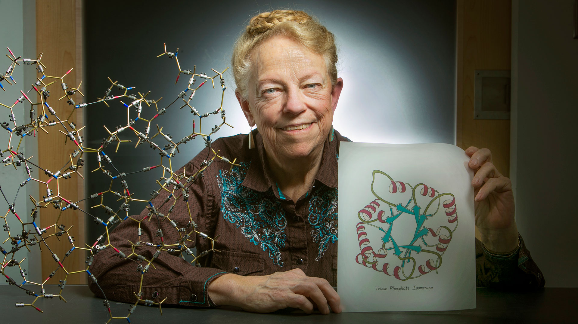
In almost every scientific paper that describes proteins you’ll find Duke biochemist Jane Richardson’s handiwork. The images, commonly referred to as “ribbon diagrams,” are basic to the language of protein science. Ranging in likeness from a flapping kite tail to a tight coil of crimped gift ribbon, these carefully etched diagrams invented by Richardson have served for many years as a primary way that scientists can describe what they see in their data.
As the elegance and complexity of proteins became more evident, scientists needed something better than wire models to articulate the subtleties they were identifying, especially in print journals. Several people had published schematic drawings of individual proteins, but each used different conventions and viewpoints. For a major review article comparing folds and details of all the known structures, Jane Richardson needed to develop a good, consistent system. She picked up a pencil and began to try drawing smooth ribbons over the atoms in early, crude computer-graphics printouts. It was promising, but hard to correctly convey the proteins in three dimensions.
“I’m not an artist and I’m not very good at drawing anything else,” she said. “It took a very long time to figure this out, how you can really show the 3D relationships on a 2D page, and also keep them accurate to the experimental atom coordinates.”